<I>Pomatoceros triqueter</I>, the keel worm
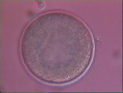
1 cell zygote
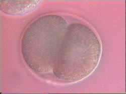
2 cell embryo
Sperm and eggs are readily isolated from the adult (video 1 below) males and females of this species. Upon mixing the gametes together embryogenesis can be followed through to hatching of the larva. (Two movies of the larva 24 (video 2) and 48 (video 3) hours post fertilization are accessible). The eggs appear to be sensitive to light which has made recording embryogenesis difficult. Only single rounds of cell division (video 4) had been captured until our 2012 visit.
A recording from fertilization through to hatching (video 5) has now been made at low magnification with low light levels. The eggs are 75 microns in diameter. The first 2 hrs 40 mins after fertilization are presented in 1 min 36 secs at 100x real time, and show initial cell divisions with 2 cell and 4 cell stages clearly visible approximately 1 minute into the movie. The next 9 hrs of development are shown in 1 min 21 secs at 400x real time. Some initial small scale movements of the embryos had not been appreciated before this recording because they are so slow in real time. The next 6 minutes of the recording are presented in 18 secs at 20x real time. The movie finishes with 15 seconds of real time to show the speed that the embryos really are moving at just before hatching, with one embryo moving from the centre to off the field of view. The original movie that this presentation has been edited from was captured by Jack Baddams, Shannon Evans, Becky Holdsworth and Nick Rushton. Low magnification with low light levels was needed because of the apparently extreme light sensitivity of this species' embryos, and consequently the resolution is also low.
Keel worm development was first studied on this course at Dale by Victoria Nichols, Ben Wardle, Sam Welply and Caroline Tibbits in Sep.'94.